Cyst Dog: What Skin Cysts Look Like and When to Get Help
A cyst on a dog is one of the most common skin findings vets see, and most of them are benign. That said, not every lump under the skin is a cyst, and the only way to know for certain is through a veterinary examination, often with a fine needle aspirate or biopsy. What does a cyst look like on a dog is a reasonable thing to want to understand before your vet appointment, because describing the lump accurately helps the vet prioritize the exam.
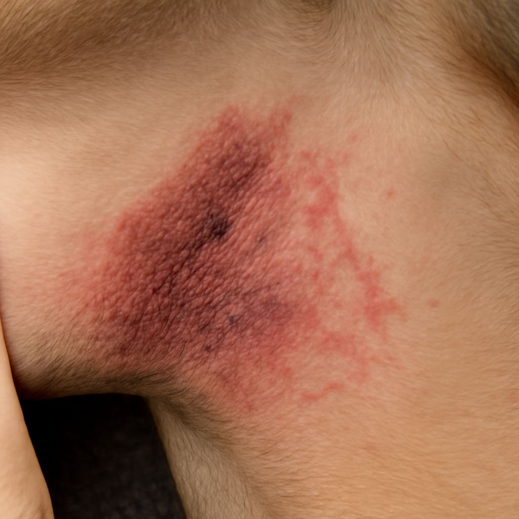
Dog skin cyst is the general term for a closed sac filled with fluid, dead skin cells, or semi-solid material. A fluid filled cyst on dog skin feels soft and movable, slightly like a water balloon under the skin. An oozing cyst on dog skin has ruptured or is draining, which means the internal pressure was high enough to breach the skin surface. Rupture reduces short-term pressure but does not resolve the cyst; the sac often refills or becomes infected if not treated.
What Does a Cyst Look Like on a Dog?
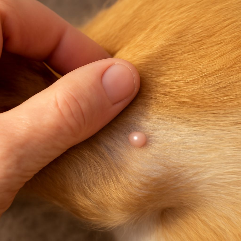
Most skin cysts appear as smooth, dome-shaped lumps that move freely when pressed. The overlying skin looks normal in early-stage cysts. As the cyst grows or fills with material, the skin above it may thin or develop a small dark pore at the center. Sebaceous cysts, the most common type, contain a white or yellowish waxy material made from sebum. Follicular cysts arise from blocked hair follicles and can grow quite large before owners notice them.
A fluid filled cyst on a dog feels distinctly different from a firm lipoma or a lymph node enlargement. Lipomas feel soft but solid, while cysts have that characteristic fluctuant quality. Location matters too: cysts develop anywhere on the body but appear most often on the back, neck, head, and legs. A cyst on the eyelid margin, called a meibomian gland cyst, is small and grows along the lid edge and may cause eye irritation if it contacts the cornea.
When an Oozing Cyst Needs Veterinary Attention
An oozing cyst on a dog becomes a concern when the discharge is bloody, foul-smelling, or accompanied by redness and swelling in the surrounding tissue. These signs suggest secondary infection. A dog skin cyst that has ruptured should be cleaned gently with saline, kept from licking with an e-collar, and examined by a vet within 24 to 48 hours.
Do not attempt to squeeze or pop a cyst at home. Squeezing forces material deeper into surrounding tissue, causing inflammation, pain, and a higher likelihood of infection. If the cyst wall ruptures internally from pressure, the result is a sterile abscess that is more complicated to treat than the original cyst. A veterinarian may drain the cyst, surgically remove it with the intact wall, or recommend monitoring if it is small and stable.
Treatment Options for Dog Skin Cysts
Small, non-infected cysts that are not bothering the dog are often monitored rather than removed immediately. Any cyst that is growing rapidly, located near the eye or a joint, or repeatedly rupturing should be surgically excised. Complete removal of the cyst wall is key: leaving the wall behind allows the cyst to regrow. Your vet may send the removed tissue for histopathology to confirm it is benign. For cysts with infection, a course of antibiotics controls the bacterial component before or after removal. Follow all post-operative care instructions and keep the incision site clean and dry until the vet confirms healing is complete.