Pictures of Dog Tumors and Cysts: Visual Guide to Common Growths
Owners searching for pictures of dog tumors and cysts often hope a quick visual comparison will tell them whether their dog’s lump is serious. The truth is that even experienced veterinarians cannot definitively diagnose a growth by appearance alone — fine needle aspiration or biopsy is required. That said, understanding visual differences between common growth types helps owners communicate more accurately and decide how urgently to seek care.
Dog cyst vs tumor is one of the most common diagnostic questions raised in veterinary dermatology consultations. Cysts and tumors genuinely do look different in many cases, but overlapping appearances make visual-only assessment unreliable. Dog ear tumor pictures, dog cyst pictures, and dog tumor pictures serve a useful purpose when used to prepare for a veterinary visit rather than replace one.
Dog Cyst vs Tumor: Key Visual and Physical Differences
What Dog Cyst Pictures Typically Show
Cysts in dogs appear as round or oval structures beneath the skin that feel smooth, moveable, and somewhat fluid-filled under finger pressure. Sebaceous cysts — the most common type — are benign and arise from blocked hair follicles. Their surface is often pale or slightly yellowish, and a whitish, pasty discharge may be visible if the cyst ruptures.
Follicular cysts, epidermoid cysts, and apocrine gland cysts follow a similar visual pattern: dome-shaped, clearly defined border, non-attached to underlying tissue. In photographs, these canine skin cysts typically appear as soft, raised bumps without surface ulceration or irregular edges. They do not typically change rapidly in size.
What Dog Tumor Pictures Reveal
Canine tumor photographs present more visual variety because tumors span dozens of types. Lipomas (benign fat tumors) look soft and well-defined, similar to cysts, but feel softer and are not fluid-filled. Mast cell tumors are more variable — they can resemble inflamed insect bites, ulcerated nodules, or firm subcutaneous masses, and their appearance changes rapidly.
Malignant growth images often show irregular borders, ulcerated or bleeding surfaces, and rapid size change over days to weeks. Melanomas around the mouth or nailbed appear dark and pigmented. These visual features are warning signs that warrant immediate evaluation, not watchful waiting.
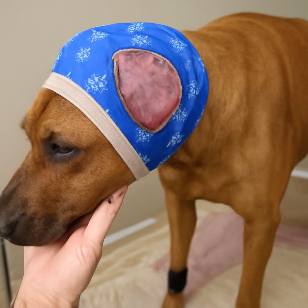
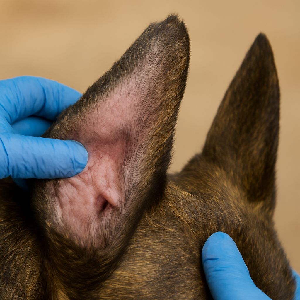
Ear-Specific Growths and Their Significance
Growths inside the ear canal or on the external pinna carry location-specific risks. Ear canal tumors can obstruct hearing, cause chronic infection, and — in malignant cases — invade surrounding tissue. Photographs of canine aural masses show pink, fleshy polyps and firm nodular outgrowths that differ markedly from normal ear canal tissue.
Ceruminous gland adenocarcinomas, the most common malignant ear canal tumor in dogs, appear as ulcerated, irregular masses within the canal. Benign inflammatory polyps look smoother and pale pink. Any growth within the ear canal, regardless of visual appearance, requires otoscopic evaluation — home assessment from ear canal images is not reliable.
When Visual Identification Is Not Enough
Visual reference images have genuine value for helping owners describe a growth to a veterinarian and track changes between appointments. They are not a substitute for cytology. A lump that looks benign in comparison to reference photos has still produced diagnostic surprises in clinical practice.
Document any growth with consistent photographs in good lighting, note the date, and record changes in size, color, or texture over time. Bring this record to the veterinary appointment. Any growth that ulcerates, bleeds, doubles in size within four weeks, or is accompanied by systemic symptoms such as weight loss or lethargy requires priority evaluation rather than deferred monitoring.
Key takeaways: Dog cyst pictures and dog tumor pictures help owners recognize patterns but cannot replace professional cytological or histological diagnosis. Visual features like irregular borders, ulceration, and rapid growth are red flags requiring urgent veterinary attention. Photograph and document any new growth and bring the record to your next appointment.