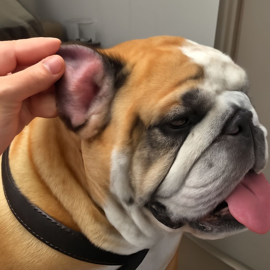
A dog ear hematoma looks alarming, but it is not immediately life-threatening. What you’re seeing is blood that has pooled between the cartilage and skin of the ear flap, usually after trauma or repeated scratching. Ear hematoma dog cases are common in floppy-eared breeds like Cocker Spaniels, Basset Hounds, and Golden Retrievers, though any dog can develop one. The swelling can appear overnight and reach the size of a golf ball, which is why owners often panic when they first notice it.
The key misunderstanding is that a hematoma dog ear condition will resolve on its own if left alone. It can, technically, but the healing process takes weeks and almost always results in a thickened, cauliflower-shaped ear from scar tissue. A hematoma in dog ear tissue that isn’t treated promptly also tends to be quite uncomfortable, and dogs often continue scratching, making it worse. Dog hematoma ear cases rarely need emergency care, but they do need a vet visit within a day or two of appearance.
What Causes an Ear Hematoma in Dogs
The Role of Head Shaking and Scratching Ear hematomas don’t appear randomly. They almost always follow repeated head shaking or ear scratching intense enough to rupture small blood vessels inside the ear flap. The underlying cause is usually an ear infection, ear mites, or allergies, all of which make the ear itchy enough to trigger that level of scratching. A hematoma dog ear is a symptom, not a standalone problem. Treating only the hematoma without identifying and resolving the underlying ear issue means the dog is likely to develop another one.
Confirming the Diagnosis
A vet can confirm a dog ear hematoma by physical examination. In some cases, a fine needle aspirate is done to confirm the contents are blood rather than serum or infection. This also guides treatment planning.
Treatment Options
The standard treatment for a hematoma in dog ear cases is surgical drainage. The vet makes a small incision, drains the collected blood, and places sutures through the ear flap in a quilting pattern to prevent fluid from re-accumulating in the empty pocket. The sutures stay in for two to three weeks. This approach has a high success rate when the underlying cause is also addressed.
A less invasive option is needle aspiration, where the fluid is drawn out with a syringe. This is faster and requires no anesthesia, but has a higher recurrence rate because the pocket can refill without the compression sutures to hold the tissue layers together. Some dog hematoma ear cases are also managed with a small drain placed in the ear that allows fluid to exit over several weeks while the ear heals around it.
Steroid injections are sometimes used for very small hematomas, particularly in dogs that are poor surgical candidates due to age or health status. Results are less predictable than surgical repair.
Recovery and Preventing Recurrence
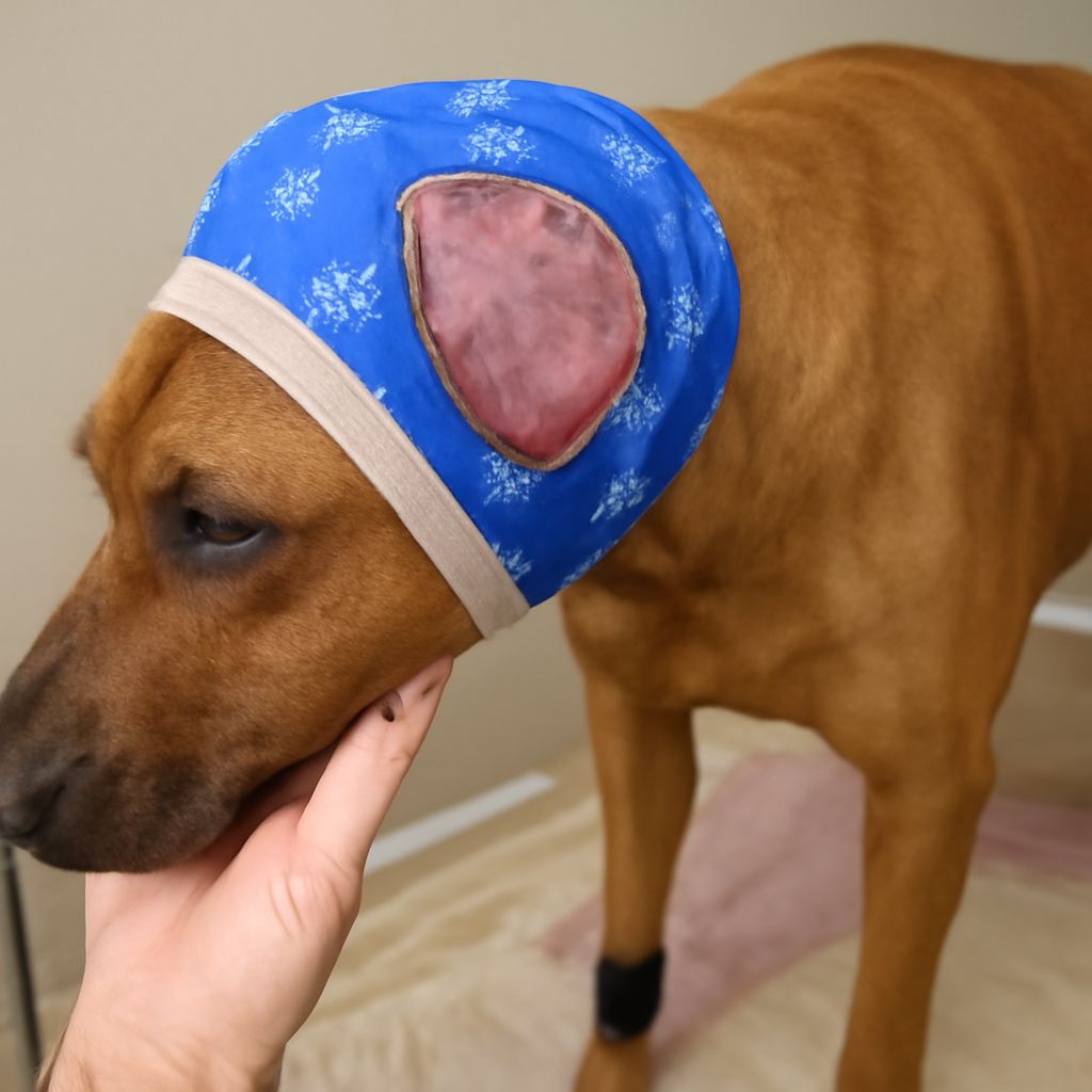
After surgery, the dog typically wears an e-collar to prevent scratching. The ear may look swollen and feel firm during healing. Final appearance depends on how long the hematoma was present before treatment and how the scar tissue forms. Early treatment gives the best cosmetic outcome.
Preventing a second ear hematoma dog case means treating the root cause. A vet will check for ear infection, mites, or allergic skin disease and address those specifically. Regular ear cleaning with a vet-recommended solution, especially in floppy-eared breeds, reduces the risk of the infections that lead to hematomas.