A corneal ulcer dog owners notice most often appears as a cloudy or bluish spot on the eye, sometimes with squinting or tearing. Many people assume a dog with a sore eye just has minor irritation, but an eye ulcer dog develops from a real wound on the surface of the cornea and needs veterinary attention. Delaying treatment makes the ulcer deeper and more difficult to heal. White dog bone chews and antlers are sometimes linked to eye injuries when a splinter or hard fragment scratches the eye during aggressive chewing. Dog blood types do not directly affect ulcer risk, but knowing your dog’s overall health profile helps your vet plan safe anesthesia if a procedure is needed. A dog corneal ulcer that is treated within 24 to 48 hours of onset heals much faster than one left alone for days.
Not every cloudy eye signals ulceration. Cataracts, nuclear sclerosis, and dry eye produce similar appearances. The difference is that corneal ulcers cause pain, so the dog squints, rubs at the face, and avoids bright light. If you see those signs together, schedule a vet visit the same day rather than waiting to see whether it clears on its own.
What Causes a Corneal Ulcer in Dogs
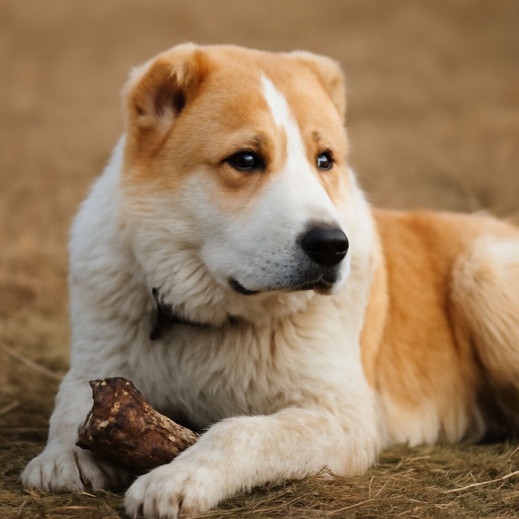
Common Injuries and Scratches Most ulcers start with physical trauma. A branch, another dog’s claw, or a cat scratch across the eye surface breaks the epithelium and starts the wound. Brachycephalic breeds like French Bulldogs, Pugs, and Boston Terriers have prominent eyes that protrude beyond the bony orbit, making them far more vulnerable to scrapes during play or rough contact with vegetation. Dogs that dig enthusiastically in dirt or sand regularly introduce small particles to the eye surface.
Hard chew items are another underappreciated cause. When a dog grips a bone or antler and gnaws intensely, small chips can fly toward the face. An eye ulcer in dogs can begin from a fragment as tiny as a grain of sand embedding in or grazing the cornea.
Chemical or Environmental Irritants
Shampoos, soaps, and cleaning sprays that splash into the eye during bath time strip the protective tear film and can initiate a superficial ulceration. Chlorine from pools and saltwater at the beach produce similar low-level chemical irritation. Chronic dry eye, where tear production is insufficient, leaves the corneal surface fragile and prone to ulcers even without a direct blow.
Symptoms That Point to an Eye Ulcer
Pain and Discharge Signs
The clearest early warning is squinting, also called blepharospasm. The dog holds the affected eye partially or fully closed and resists having it touched. Watery discharge is common in early-stage ulcers, while thicker yellow or green discharge signals secondary bacterial infection taking hold. Rubbing the eye against furniture or the floor worsens the injury and can cause a shallow ulcer to become a deep stromal one within hours.
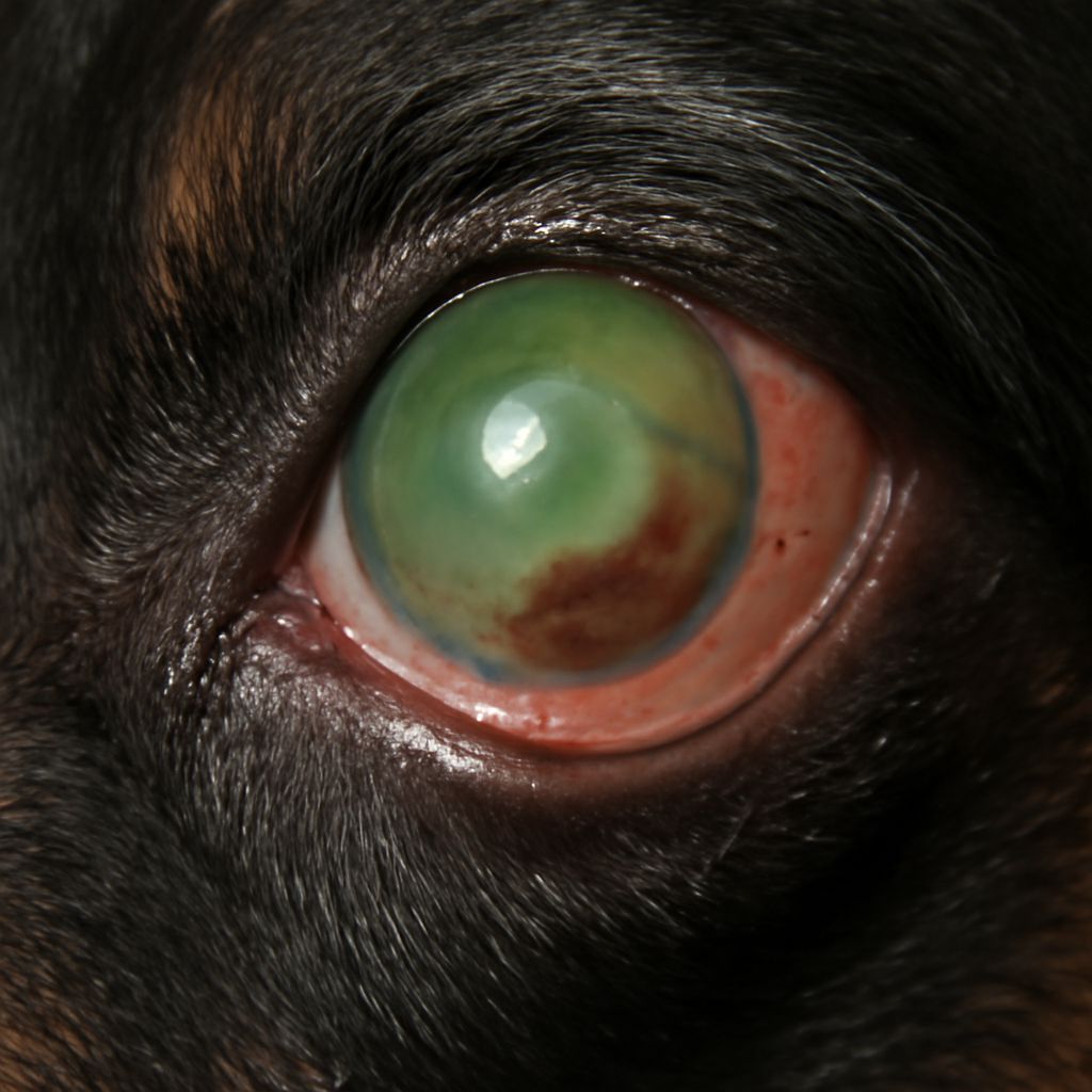
Cloudiness and Vision Changes As the immune system responds to corneal injury, white blood cells migrate into the tissue and produce visible haze or opacity. A dog corneal ulcer that has progressed to stromal involvement shows a clearly white or gray patch in the center of the eye. In advanced cases the cornea develops new blood vessels growing inward from the edges, a process called vascularization, which is the body’s attempt to deliver healing cells to damaged tissue.
How Dog Corneal Ulcers Are Diagnosed and Treated
Vet Diagnostic Methods
Vets use fluorescein stain, an orange dye applied as drops, to confirm ulceration. The stain adheres to exposed stromal tissue and glows green under a blue light, making even shallow defects visible. This is painless and gives an accurate picture of ulcer depth and boundaries within minutes. A Schirmer tear test rules out dry eye as an underlying cause, and tonometry checks intraocular pressure to ensure no secondary glaucoma has developed.
Medications and Recovery Steps
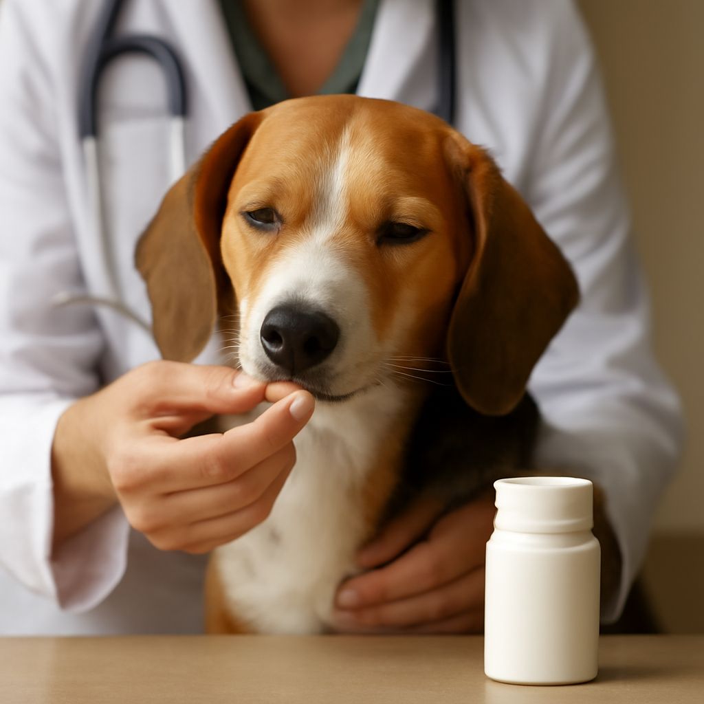
Treatment depends on ulcer depth. Superficial ulcers respond to antibiotic eye drops or ointment, applied three to four times daily, combined with an Elizabethan collar to prevent self-trauma. Atropine drops widen the pupil and reduce painful ciliary muscle spasm. Most simple corneal injuries in dogs heal within five to seven days. Deep or nonhealing ulcers require surgical intervention, such as a grid keratotomy or conjunctival graft, performed by a veterinary ophthalmologist. Follow every recheck appointment without skipping, since a healing ulcer can reverse rapidly if the dog rubs at it once.